
Free ATI TEAS 7 Science guide
October 2026 - Cardiovascular System Study Guide for nursing students in
The cardiovascular system moves blood in two loops through a four-chambered heart: the right side sends oxygen-poor blood to the lungs, the left side sends oxygen-rich blood to the body. Get the order of the chambers and valves fixed in your memory and most questions on this topic answer themselves.
The path of blood through the heart
This single sequence is the most heavily tested thing in the topic. Blood arrives from the body deoxygenated, is sent to the lungs, comes back oxygenated, and is then pushed out to the body again — and it passes a valve on the way out of every chamber.
- Body
- Vena cava
- Right atrium
- Tricuspid valve
- Right ventricle
- Pulmonary valve
- Pulmonary artery
- Lungs
- Pulmonary vein
- Left atrium
- Mitral valve
- Left ventricle
- Aortic valve
- Aorta
- Body
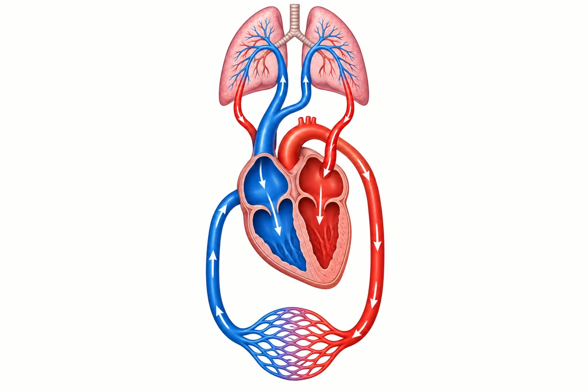
Two habits make this reliable under time. First, learn it as a loop you can start anywhere rather than a list you recite from the beginning, because questions ask what comes next, not what comes first. Second, notice the alternation: chamber, valve, chamber, valve. Atria receive, ventricles pump, and a valve sits at every exit to stop backflow.
The two atrioventricular valves — tricuspid on the right, mitral on the left — separate each atrium from the ventricle below it. The two semilunar valves — pulmonary and aortic — guard the exits from the ventricles into the great arteries.
The two circuits
Everything the heart does is one of two loops, and each loop is driven by one side of the heart.
- Pulmonary circulation — the right side of the heart to the lungs and back. It carries oxygen-poor blood out and returns oxygen-rich blood.
- Systemic circulation — the left side of the heart to the rest of the body and back. It carries oxygen-rich blood out and returns oxygen-poor blood.
- Why the left ventricle is the thickest chamber — it has to generate enough pressure to push blood through the entire body, while the right ventricle only has to reach the lungs next door. Thickness follows workload.
The naming rule is about direction, not oxygen. Arteries carry blood away from the heart and veins carry it back — which is why the pulmonary artery is the one artery carrying oxygen-poor blood, and the pulmonary vein the one vein carrying oxygen-rich blood. Questions are built on exactly this exception.
Blood vessels
Three vessel types, distinguished by direction of flow and by wall structure.
| Vessel | Direction | Structure | Job |
|---|---|---|---|
| Arteries | Away from the heart | Thick, muscular, elastic walls | Withstand high pressure from the ventricles |
| Veins | Back to the heart | Thinner walls, one-way valves | Return blood at low pressure against gravity |
| Capillaries | Between the two | One cell thick | Exchange of gases, nutrients and waste with tissue |
What blood is made of
- Red blood cells (erythrocytes) — carry oxygen bound to haemoglobin, an iron-containing protein. Too little iron means too little functional haemoglobin, which is why iron-deficiency anaemia presents as fatigue and breathlessness.
- White blood cells (leukocytes) — the immune cells. They rise in number during infection.
- Platelets (thrombocytes) — cell fragments that begin clotting at a site of injury.
- Plasma — the straw-coloured liquid, mostly water, that carries the cells along with hormones, proteins, nutrients and waste.
Blood pressure and the heartbeat
A blood pressure reading is written as systolic over diastolic — for example 118/76 mmHg. Systolic, the larger number, is the pressure while the ventricles contract. Diastolic, the smaller number, is the pressure while they relax and refill. The larger number is always on top.
The beat itself is electrical. The sinoatrial (SA) node in the right atrium fires on its own and sets the rate, which is why it is called the pacemaker. The signal spreads across the atria to the atrioventricular (AV) node, which delays it just long enough for the atria to finish emptying before the ventricles contract.
Reading a question that hides the sequence
Worked example
A clot forms in the pulmonary artery. Which chamber of the heart is immediately upstream of the blockage, and what happens to the blood behind it?
- Locate the pulmonary artery in the loop: it sits between the pulmonary valve and the lungs.
- Step back one place. The chamber before the pulmonary valve is the right ventricle.
- Ask what that blood was on its way to do. It was going to the lungs to pick up oxygen, so it is oxygen-poor.
- A blockage there means oxygen-poor blood backs up in the right ventricle and cannot reach the lungs for gas exchange.
The right ventricle. Oxygen-poor blood accumulates behind the clot and never reaches the lungs to be oxygenated.
Terms to know
- Systole
- The contraction phase, when a chamber squeezes blood out. The top number of a blood pressure reading.
- Diastole
- The relaxation phase, when a chamber refills. The bottom number of a blood pressure reading.
- Atrium
- An upper, receiving chamber. Blood arrives here from a vein.
- Ventricle
- A lower, pumping chamber. Blood leaves here through an artery.
- SA node
- The sinoatrial node, in the right atrium. It generates the heartbeat and sets the rate — the natural pacemaker.
- Haemoglobin
- The iron-containing protein in red blood cells that oxygen binds to.
- Septum
- The muscular wall dividing the right side of the heart from the left, keeping the two circuits separate.
- Pulse
- The wave of pressure felt in an artery each time the left ventricle contracts.
What the TEAS asks most
The same core ideas appear in different wording. If you can answer these without stopping to think, you have what this section requires.
Which chamber or vessel does blood reach next?
Follow the loop: vena cava, right atrium, tricuspid, right ventricle, pulmonary valve, pulmonary artery, lungs, pulmonary vein, left atrium, mitral, left ventricle, aortic valve, aorta.
Which vessel carries oxygen-poor blood away from the heart?
The pulmonary artery. It is the exception to "arteries carry oxygenated blood", because artery describes direction, not oxygen content.
Why is the wall of the left ventricle thicker than the right?
It pumps against the resistance of the whole body, while the right ventricle only pumps to the lungs.
What does the top number in a blood pressure reading measure?
Systolic pressure — the pressure in the arteries during ventricular contraction.
What structure starts each heartbeat?
The SA node in the right atrium.
Key points
- Blood makes two loops through one heart: right side to the lungs, left side to the body.
- Chamber, valve, chamber, valve — a valve sits at the exit of every chamber to prevent backflow.
- Arteries carry blood away from the heart, veins carry it back. The pulmonary vessels are the oxygen exceptions.
- The left ventricle is the thickest chamber because it pumps against the whole body.
- Systolic over diastolic: contraction over relaxation, larger number on top.
- Capillaries are one cell thick, which is what makes exchange with tissue possible.
Review quiz
5 questions on this topic, each with the reasoning worked through. Pick an answer to see how it went, or open the explanation straight away.
-
Question 1 of 5
Blood leaving the right ventricle passes through the pulmonary valve. Where does it travel next?
Show the answer and explanation
Correct answer: B. The pulmonary artery
The pulmonary valve opens into the pulmonary artery, which carries oxygen-poor blood to the lungs. The aorta is the exit from the left ventricle, not the right; the left atrium is where blood arrives after the lungs; and the vena cava delivers blood into the right atrium at the start of the loop.
-
Question 2 of 5
Which vessel carries oxygen-poor blood away from the heart?
Show the answer and explanation
Correct answer: C. The pulmonary artery
The pulmonary artery carries oxygen-poor blood from the right ventricle to the lungs. This is the trap the question is built on: artery means away from the heart, not oxygen-rich. The pulmonary vein is the mirror-image exception — a vein carrying oxygen-rich blood back to the left atrium.
-
Question 3 of 5
A patient has a blood pressure of 132/84 mmHg. What does the 84 represent?
Show the answer and explanation
Correct answer: A. Arterial pressure while the ventricles are relaxing and refilling
The lower number is diastolic pressure, measured while the ventricles relax and fill. The 132 is systolic, measured during contraction. Heart rate and stroke volume are separate measurements that a blood pressure reading does not report.
-
Question 4 of 5
A student with low dietary iron reports constant tiredness and breathlessness on stairs. Which cardiovascular component most directly explains the symptoms?
Show the answer and explanation
Correct answer: D. Red blood cells, because haemoglobin needs iron to carry oxygen
Haemoglobin is an iron-containing protein, so too little iron means red blood cells carry less oxygen and tissues run short during exertion. Platelets handle clotting, plasma volume is governed by fluid balance rather than iron, and white blood cells are immune cells — none of the three would produce this pattern of symptoms.
-
Question 5 of 5
Which feature of capillaries makes gas and nutrient exchange with body tissue possible?
Show the answer and explanation
Correct answer: B. Walls only one cell thick
Exchange happens by diffusion, and diffusion needs a very short distance to cross, so a wall one cell thick is the whole point of a capillary. Thick muscular walls describe arteries, one-way valves describe veins, and capillaries are in fact the narrowest vessels in the body, not the widest.
Put this TEAS topic into practice
Build a set on cardiovascular system and use the rationale on every question to reinforce what you just reviewed.
Keep reading
More TEAS Resources
Guides and tools across the TEAS course.
TEAS 7 Exam Prep
Format, sections, timing
TEAS 7 FAQ
Scoring, retakes, test day
Is the TEAS Hard?
An honest answer
How Long Is the TEAS 7?
209 minutes, section by section
What Is a Passing TEAS Score?
Your programme sets the bar
TEAS Reading
18 topics · 39 scored questions
TEAS Math
18 topics · 34 scored questions
TEAS Science
22 topics · 44 scored questions
TEAS English
21 topics · 33 scored questions
Free TEAS Practice Test
Sit a set before you buy
TEAS Plans & Pricing
Choose your access
Start TEAS Prep
Create your account
TEAS Study Guide
All four sections, free
8-Week Study Plan
Week-by-week schedule
TEAS Cheat Sheet
Formulas and high-yield facts
TEAS vs HESI A2
Which exam to take