Free HESI A2 Anatomy and Physiology guide
October 2026 - Anatomical Imaging Study Guide for nursing students in
Anatomical imaging turns internal structures into interpretable pictures by using different forms of energy and different viewing planes. For HESI A2 preparation, focus on what each common modality shows well, its basic mechanism and how orientation changes the view.
Core ideas to know
Learn the relationships, not a string of isolated definitions. These are the facts that let you reason through unfamiliar wording.
- Radiography uses ionising x-rays and shows dense structures such as bone strongly because they absorb more radiation.
- Computed tomography (CT) combines many x-ray measurements into cross-sectional images and therefore also uses ionising radiation.
- Magnetic resonance imaging (MRI) uses a magnetic field and radiofrequency energy, giving strong soft-tissue contrast without x-rays.
- Ultrasound forms real-time images from reflected high-frequency sound and is useful for motion and fluid-filled structures.
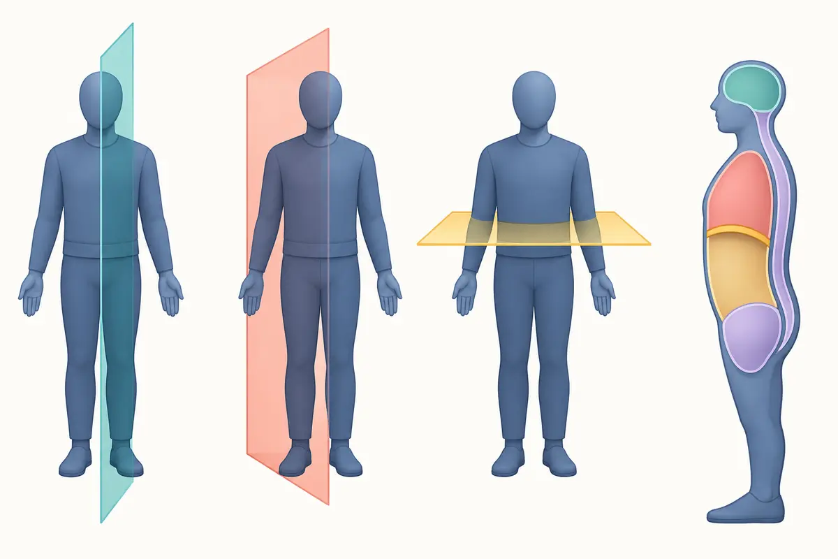
- A cross-sectional image may be sagittal, frontal or transverse, and its labels should be read before assuming orientation.
A reliable way to answer
Use the same short process every time so the wording of the question does not decide your method for you.
- Identify the modality from its stated energy source: x-ray, magnet and radio waves, or sound.
- Identify the plane and locate obvious landmarks before naming a smaller structure.
- Compare tissue density or signal within that modality rather than across unrelated image types.
- Choose the modality whose strengths match the structure or process described in the stem.
Distinctions that earn marks
| Idea | What it means | Do not confuse it with |
|---|---|---|
| CT | Cross-sectional x-ray imaging | MRI, magnetic and radiofrequency imaging |
| Ultrasound | Reflected sound with real-time motion | Radiograph, projection made with x-rays |
| Transverse image | Superior and inferior portions | Sagittal image, left and right portions |
Read every word in the stem before choosing. HESI distractors are often true statements that answer a nearby question rather than the one that was asked.
Terms to know
- Modality
- A particular imaging method and its physical mechanism.
- Contrast
- The visible difference that lets adjacent tissues be distinguished.
- Cross-section
- An image representing a slice through the body.
Key points
- Radiographs and CT use ionising x-rays; MRI and ultrasound do not.
- CT is cross-sectional, while an ordinary radiograph is a projection.
- Always establish plane and orientation before interpreting a labelled image.
Review quiz
3 questions on this topic, each with the reasoning worked through. Pick an answer to see how it went, or open the explanation straight away.
-
Question 1 of 3
Which modality uses high-frequency sound waves?
Show the answer and explanation
Correct answer: C. Ultrasound
Ultrasound sends and receives high-frequency sound waves to form an image. CT and radiography use x-rays, while MRI uses magnetic and radiofrequency energy.
-
Question 2 of 3
Why does bone usually appear prominent on a conventional radiograph?
Show the answer and explanation
Correct answer: B. Its density absorbs more x-rays than surrounding soft tissue.
Dense mineralised bone attenuates more of the x-ray beam, creating strong contrast with less-dense soft tissue. The image does not depend on bone emitting energy.
-
Question 3 of 3
Which statement correctly compares CT and MRI?
Show the answer and explanation
Correct answer: B. CT uses x-rays; MRI uses magnetic and radiofrequency energy.
CT reconstructs measurements made with x-rays, whereas MRI relies on a strong magnetic field and radiofrequency pulses. Both can display cross-sectional anatomy.
Put this HESI topic into practice
Build a set on anatomical imaging and use the rationale on every question to reinforce what you just reviewed.
Keep reading
More HESI Resources
Guides and tools across the HESI course.
HESI A2 Exam Prep
Subjects and format
HESI A2 FAQ
Scoring, retakes, test day
Which HESI Am I Taking?
A2, specialty or exit exam
Which Subjects Will I Sit?
The full Elsevier menu
HESI Math
4 topics · ratios and conversions
HESI Reading
16 topics · inference and tone
HESI Vocabulary
9 topics · affixes and word choice
HESI Grammar
7 topics · usage and punctuation
HESI Anatomy & Physiology
16 topics · one system at a time
HESI Biology
13 topics · cells and genetics
HESI Plans & Pricing
Choose your access
Start HESI Prep
Create your account
Ask Your Programme
What to confirm before you book
HESI A2 Study Guide
Six covered subjects, free
6-Week Study Plan
Week-by-week schedule
HESI Cheat Sheet
Formulas and high-yield facts
TEAS vs HESI A2
Which exam to take